Category:Molecular biology
Jump to navigation
Jump to search
branch of biology that deals with the molecular basis of biological activity | |||||
| Upload media | |||||
| Instance of | |||||
|---|---|---|---|---|---|
| Subclass of | |||||
| Part of | |||||
| Has part(s) |
| ||||
| |||||
English: Molecular biology is the study of biology at a molecular level. The field overlaps with other areas of biology, particularly genetics and biochemistry. Molecular biology chiefly concerns itself with understanding the interactions between the various systems of a cell, including the interrelationship of DNA, RNA and protein synthesis and learning how these interactions are regulated.
Subcategories
This category has the following 77 subcategories, out of 77 total.
*
B
- Bacterial one-hybrid system (13 F)
C
- Cre-Lox recombination (9 F)
D
E
F
G
- Genetic circuit (8 F)
- GUS reporter system (5 F)
H
I
M
- Media from EMBO Reports (5 F)
- Media from Molecular Brain (52 F)
- Media from Molecular Cancer (48 F)
N
O
- Open reading frames (21 F)
- Optical tweezers (90 F)
P
S
- Screening and selection (10 F)
- Site-directed mutagenesis (52 F)
- Southern blot (13 F)
T
- Two-hybrid system techniques (29 F)
W
- WikiPathways (39 F)
Pages in category "Molecular biology"
This category contains only the following page.
Media in category "Molecular biology"
The following 200 files are in this category, out of 303 total.
(previous page) (next page)-
(zh)2A peptide Working Mechanism.jpg 593 × 493; 125 KB
-
2A peptide Working Mechanism.jpg 593 × 493; 129 KB
-
41598 2018 20107 Fig3 HTML.jpg 660 × 315; 167 KB
-
4nqo1.gif 521 × 119; 6 KB
-
4nqo2.png 784 × 194; 60 KB
-
Acquaporina subunità porocanale.png 986 × 492; 27 KB
-
Affinity Chromatography.jpg 580 × 800; 161 KB
-
Affymetrix GeneChip.jpg 750 × 600; 312 KB
-
Ahelix-FMO-Facio.jpg 1,073 × 821; 105 KB
-
Annotated structure of eRF1.jpg 600 × 361; 81 KB
-
Apotosis.jpg 1,302 × 520; 157 KB
-
Application of MNAse.png 959 × 540; 194 KB
-
Arnmensajero1.png 283 × 212; 28 KB
-
ASO dot blot inverso.png 1,310 × 786; 42 KB
-
ASO dot blot.png 1,304 × 864; 38 KB
-
Balizas moleculares.png 960 × 720; 11 KB
-
Bilal Djeghout Laboratory.jpg 1,966 × 2,048; 361 KB
-
Binding.png 888 × 572; 22 KB
-
Biological information flow.tif 1,150 × 461; 2.57 MB
-
Biological Safety Cabinet (Class II, Type A2) Front view.jpg 1,469 × 1,102; 420 KB
-
Biological Safety Cabinet (Class II, Type A2) Side view.jpg 1,469 × 1,102; 503 KB
-
Biological Safety Cabinet (Class II, Type A2) Telstar front view.jpg 2,048 × 1,536; 731 KB
-
Canonical and non-canonical Wnt signaling pathways.png 3,620 × 2,214; 1.06 MB
-
Cascada JNK.jpg 2,001 × 1,501; 194 KB
-
Cassettes.png 960 × 720; 42 KB
-
CDNA-library-amplification.png 2,955 × 1,419; 159 KB
-
Central Dogma Model.png 800 × 557; 53 KB
-
Choosing a cloning method.png 2,625 × 1,425; 144 KB
-
Chromatogramme.jpg 583 × 768; 51 KB
-
ColonneADN.JPG 153 × 359; 4 KB
-
ColonneElution.JPG 153 × 359; 6 KB
-
ConceptLarge.jpg 97 × 380; 28 KB
-
Condensation3.png 2,852 × 791; 279 KB
-
COREcassette.PNG 960 × 720; 43 KB
-
Courtney 2008.jpg 786 × 685; 52 KB
-
CreLoxP.jpg 320 × 316; 13 KB
-
Crystal-by-example-illustrating-how-a-helix-might-be-formed.jpg 600 × 514; 82 KB
-
CSIRO ScienceImage 2082 Cell Culture.jpg 2,657 × 1,976; 5.15 MB
-
Ctd role .png 278 × 478; 22 KB
-
Deconvoluted ESMS.jpg 1,648 × 1,123; 71 KB
-
Dien bien nhanh.JPG 478 × 709; 64 KB
-
Dieu hoa di hinh lap the-01.png 9,542 × 6,723; 652 KB
-
DL20221201 oomikad.tif 4,000 × 2,250; 720 KB
-
DL20221209 2nd-messenger.tif 945 × 1,654; 219 KB
-
DL20230220 Fsk effect CHO H188.tif 3,558 × 1,902; 138 KB
-
DNA repair.png 1,080 × 1,350; 627 KB
-
DNA-Leiter.jpg 404 × 802; 55 KB
-
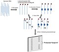
DNaseI footprint.png 162 × 626; 79 KB
-
Donning.jpg 1,102 × 1,469; 440 KB
-
Droga alternatywna.png 759 × 350; 66 KB
-
Droga klasyczna.png 725 × 405; 74 KB
-
Ecoli human compare.jpg 675 × 450; 51 KB
-
Electronic access control (BSL3 Lab) using magnetic swipe card.jpg 2,048 × 1,536; 702 KB
-
Electronic access control (BSL3 Lab) using personal identification number (PIN).jpg 2,048 × 1,536; 732 KB
-
Electronic access control (BSL3 Lab).jpg 2,048 × 1,536; 725 KB
-
Epissage.png 565 × 222; 10 KB
-
Epitelo-mezenchymální tranzice.gif 822 × 214; 36 KB
-
Erkennungssequenzen von Restriktionsenzymen.jpg 764 × 331; 22 KB
-
EsquemaBiologiaMolecular.png 576 × 354; 599 KB
-
Estructura HERC1.jpg 1,585 × 1,665; 244 KB
-
Estructura ORC.png 399 × 216; 21 KB
-
Experimental technique Dip-c.jpg 1,771 × 338; 101 KB
-
Experimento-pulso-caza.jpg 500 × 272; 39 KB
-
Extraction Chamber of Molecular Laboratory.jpg 4,000 × 2,250; 2.39 MB
-
Extrapolation based Molecular Systems biology GRana 09.jpg 960 × 720; 73 KB
-
Extrapolation based Molecular Systems biology Rana 09.jpg 960 × 720; 73 KB
-
Extrapolation Based Molecular Systems Biology Rana CWRU.tiff 960 × 720; 589 KB
-
Faire scheme.png 3,000 × 2,300; 1.78 MB
-
Fatty Acid Transport Mechanism.png 1,190 × 872; 534 KB
-
Fedg.png 1,086 × 817; 363 KB
-
Fig2.Recombination patterns.png 960 × 720; 12 KB
-
Figura Wiki.png 720 × 504; 206 KB
-
Figure 1 NAPPA.png 1,363 × 470; 69 KB
-
Figure 2 PISA.png 1,315 × 374; 61 KB
-
Figure 3 puromycin2.png 1,415 × 469; 93 KB
-
Figure 4 nano well.png 1,390 × 403; 62 KB
-
Figure 5 DAPA.png 1,392 × 628; 60 KB
-
Figure final 3.jpg 3,071 × 2,457; 491 KB
-
Finite Element Model.jpg 793 × 634; 41 KB
-
Finite model.jpg 1,050 × 734; 53 KB
-
Fish Egg Diagram (1).jpg 960 × 720; 38 KB
-
Fish Egg.jpg 960 × 720; 37 KB
-
FlAsh Protein Modification.png 2,161 × 676; 110 KB
-
Functional Cloning.png 5,500 × 800; 611 KB
-
GelDoc DNA gel electrophoresis photograph stained with ethidium bromide 01.jpg 937 × 1,034; 333 KB
-
GelDoc DNA gel electrophoresis photograph stained with ethidium bromide 03.jpg 737 × 1,045; 200 KB
-
GelDoc DNA gel electrophoresis photograph stained with ethidium bromide 05.jpg 1,498 × 1,477; 528 KB
-
GelDoc DNA gel electrophoresis photograph stained with ethidium bromide 06.jpg 1,207 × 533; 299 KB
-
GelDoc DNA gel electrophoresis photograph stained with ethidium bromide 07.jpg 1,440 × 625; 227 KB
-
Genequant.jpg 1,536 × 2,048; 226 KB
-
GESTALT workflow.png 1,436 × 769; 107 KB
-
GolgiTethersc.jpg 390 × 233; 28 KB
-
Gpi synthesis.jpg 4,187 × 3,455; 1.79 MB
-
Grafica de la PCR.jpg 528 × 600; 77 KB
-
Heavy Metals vs REE vs Plant Molecular Biology.png 876 × 526; 51 KB
-
HIVE Annotation Mapper Computation.png 927 × 885; 130 KB
-
HIVE Heptagon Computation.png 1,034 × 1,161; 252 KB
-
HIVE Hexagon Computation.png 1,015 × 850; 185 KB
-
HIVE Hexahedron Computation.png 969 × 1,053; 221 KB
-
HIVE IDBA-UD Computation.png 1,102 × 885; 151 KB
-
HIVE MAFFT Computation.png 1,075 × 728; 174 KB
-
HIVE Velvet Computation.png 1,102 × 696; 121 KB
-
Hmm necleotides 2.pdf 856 × 881; 49 KB
-
Hmm nucleotide seq.pdf 1,354 × 137; 18 KB
-
Human genome to genes zh.png 1,454 × 866; 386 KB
-
Hydrophobic Mismatch.JPG 936 × 995; 164 KB
-
Hypothesis Scarano-etal.1967.png 567 × 433; 19 KB
-
Hêlicaza Helicase.png 150 × 448; 19 KB
-
Hêlicaza tác động Helicase in action.png 200 × 448; 31 KB
-
Initial model of chlororespiration.jpg 797 × 556; 29 KB
-
Isoforms and sequence.png 676 × 620; 44 KB
-
Jeewanu globules - Electron Imaging.png 342 × 446; 122 KB
-
Journal.pone.0001604.g001 small.jpg 597 × 144; 26 KB
-
Kimura three parameter substitution model.png 401 × 409; 29 KB
-
Kimura two parameter substitution model.png 401 × 409; 28 KB
-
Klonierung2.png 624 × 239; 16 KB
-
La technologie BRET.png 1,022 × 1,162; 49 KB
-
Label-free Localisation Microscopy SPDM - Super Resolution Microscopy Christoph Cremer.jpg 2,012 × 1,280; 1.55 MB
-
Latest understanding of chlororespiration (2002).jpg 937 × 527; 31 KB
-
Lentiviral vector.png 961 × 467; 126 KB
-
LH2 side.jpg 1,048 × 904; 151 KB
-
Lipofección.png 509 × 506; 141 KB
-
Mattress model.JPG 921 × 729; 92 KB
-
MCR1erythro4TC.jpg 564 × 304; 41 KB
-
Mechanism of Nonsense Mediated Decay.jpg 631 × 1,155; 150 KB
-
Mediator4TC.jpg 357 × 289; 41 KB
-
MediatorPolII4TC.jpg 326 × 224; 34 KB
-
MediatorPolTF4TC.jpg 603 × 270; 69 KB
-
MediatorSpline4TC.jpg 194 × 240; 21 KB
-
MediatorStrucMod4TC.jpg 271 × 168; 21 KB
-
Membrane lipids.png 579 × 607; 10 KB
-
Membrane potential development.jpg 1,619 × 518; 79 KB
-
Membrane potential ions (id).jpg 550 × 400; 186 KB
-
MirrorImagePhageDisplay.png 1,775 × 691; 150 KB
-
MNAse based sequencing.png 360 × 622; 94 KB
-
MNase based sequencing.png 360 × 622; 94 KB
-
Mnase image.png 360 × 622; 84 KB
-
Molecular response after nerve injury.pdf 962 × 806; 243 KB
-
Molecular response after nerve injury.png 1,926 × 1,613; 550 KB
-
Musclon2.jpg 556 × 442; 29 KB
-
MutHPvuII.png 1,075 × 498; 467 KB
-
Na-K pump cycle.jpg 800 × 457; 108 KB
-
Na-K pump cycle.png 800 × 457; 156 KB
-
NASBA fase 1.jpg 1,508 × 929; 69 KB
-
NASBA fase 2.jpg 1,495 × 1,135; 115 KB
-
Nearest-Neighbor-seq-freq XpY.png 1,665 × 886; 121 KB
-
Neocentromere Formation .jpg 2,917 × 3,508; 1.71 MB
-
Neubauer improved with cells.jpg 1,280 × 960; 669 KB
-
Neural progenitor cells.png 3,893 × 939; 637 KB
-
Neuraminidase2.jpg 785 × 662; 349 KB
-
NLRP3.png 1,460 × 976; 645 KB
-
NMD - Nonsense-mediated decay.png 7,292 × 4,965; 709 KB
-
Nrm2503-f2.jpg 655 × 440; 118 KB
-
Nuclear integrity and genome stability in normal and HGPS cells.jpg 600 × 723; 64 KB
-
Outline of no-SCAR recombineering methods final 2.png 2,400 × 2,810; 25.74 MB
-
PAPRs in use 01.jpg 1,280 × 853; 100 KB
-
Parik1.jpg 680 × 552; 65 KB
-
Parik2.1.jpeg 388 × 636; 29 KB
-
Parik3.jpeg 623 × 642; 70 KB
-
Parik4.jpg 706 × 334; 55 KB
-
Pathways of glucolysis.png 1,366 × 768; 90 KB
-
PCR es.png 300 × 675; 26 KB
-
Penetrance.pdf 1,239 × 1,752; 51 KB
-
Penetrance1.0.pdf 1,752 × 1,239; 49 KB
-
Penetrance2.pdf 1,239 × 1,752; 56 KB
-
PenetranceVE.pdf 1,752 × 1,239; 56 KB
-
Perfil de exitación y emision de DAPI.jpg 252 × 220; 13 KB
-
Peroxisome Dynamics-Molecular Players,Mechanisms, and (Dys)functions.pdf 1,250 × 1,650, 25 pages; 375 KB
-
PETworkflow.png 1,061 × 797; 75 KB
-
PGEX-3X cloning vector.png 356 × 354; 10 KB
-
Philadelphia chromosome detection.jpg 563 × 669; 133 KB
-
Physiology.. .png 427 × 387; 25 KB
-
Pipette tip over tube.jpg 2,361 × 3,305; 623 KB
-
Piskacek TF1.jpg 8,080 × 3,456; 1.57 MB
-
PL Mykowirusy – wirusy infekujące grzyby J. Kamiński.pdf 1,239 × 1,754, 56 pages; 1.82 MB
-
PL Wstępna charakterystyka bakteriofaga Serratia φOS10 J. Kamiński.pdf 1,239 × 1,752, 101 pages; 2.13 MB
-
Plant cell structure svg vacuole (id).jpg 649 × 475; 156 KB
-
Plant-cell-sucrose-gradient-fractions.jpg 2,988 × 5,312; 1.74 MB
-
Polysomesleft.jpg 402 × 518; 45 KB
-
PomBase infographic.jpg 1,591 × 2,250; 608 KB
-
Post063a - Flickr - NOAA Photo Library.jpg 1,800 × 1,200; 2.1 MB
-
Powerlaw HI II 14.png 536 × 391; 24 KB
-
Principle of competent cell preparation 1.png 2,149 × 1,627; 421 KB
-
Principle of competent cell preparation 2.png 2,154 × 1,627; 436 KB
-
Proces de la PCR.jpg 300 × 675; 81 KB
-
PromotorsK01589 logo 1.pdf 1,050 × 1,050; 51 KB
-
Protein co-localization on microtubules.png 2,160 × 2,160; 8.09 MB
-
Proton trap.jpg 690 × 409; 60 KB
-
Proves de la TaqMan.jpg 650 × 167; 23 KB
-
ProximityAssay14TC.jpg 269 × 92; 8 KB
-
ProximityAssay24TC.jpg 270 × 231; 15 KB
-
ProximityAssay34TC.jpg 269 × 229; 13 KB


