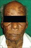
Syphilis of Fungal world: Novel Skin Manifestations of Histoplasmosis in an Immunocompetent Host
- PMID: 23248386
- PMCID: PMC3519275
- DOI: 10.4103/0019-5154.103089
Syphilis of Fungal world: Novel Skin Manifestations of Histoplasmosis in an Immunocompetent Host
Abstract
A case of chronic disseminated cutaneous histoplasmosis with unusual skin manifestations in an immunocompetent host is reported. Presence of cutaneous ulcers, linear erythematous plaques, skin coloured atrophic plaques and recurrent self-limiting oral ulcers in a single patient has not been documented in literature so far. Diagnosis was established by identifying small intracellular yeast-like cells of Histoplasma in tissue smear and skin biopsy. Leishman stained tissue smear proves to be an easy and simple procedure for diagnosis of histoplasmosis.
Keywords: Histoplasmosis; itraconazole; tissue smear.
Conflict of interest statement
Figures


Similar articles
-
Disseminated cutaneous histoplasmosis in an immunocompetent adult.Indian J Dermatol. 2012 May;57(3):206-9. doi: 10.4103/0019-5154.96194. Indian J Dermatol. 2012. PMID: 22707773 Free PMC article.
-
Unusual presentation of disseminated histoplasmosis in an immunocompetent patient.Diagn Cytopathol. 2017 Sep;45(9):848-850. doi: 10.1002/dc.23742. Epub 2017 May 4. Diagn Cytopathol. 2017. PMID: 28474430
-
Disseminated histoplasmosis presenting as pyoderma gangrenosum-like lesions in a patient with acquired immunodeficiency syndrome.Int J Dermatol. 2001 Aug;40(8):518-21. doi: 10.1046/j.1365-4362.2001.01254.x. Int J Dermatol. 2001. PMID: 11703524
-
An Unusual Presentation of Disseminated Histoplasmosis: Case Report and Review of Pediatric Immunocompetent Patients from India.Mycopathologia. 2015 Dec;180(5-6):359-64. doi: 10.1007/s11046-015-9917-y. Epub 2015 Jul 1. Mycopathologia. 2015. PMID: 26126955 Review.
-
[Primary cutaneous histoplasmosis: case report on an immunocompetent patient and review of the literature].Rev Soc Bras Med Trop. 2008 Nov-Dec;41(6):680-2. doi: 10.1590/s0037-86822008000600024. Rev Soc Bras Med Trop. 2008. PMID: 19142453 Review. Portuguese.
Cited by
-
Hiding in plain sight: a case of chronic disseminated histoplasmosis with central nervous system involvement.BMJ Case Rep. 2017 Jul 6;2017:bcr2017220476. doi: 10.1136/bcr-2017-220476. BMJ Case Rep. 2017. PMID: 28687695 Free PMC article.
References
-
- Hay RJ. Deep fungal infections. In: Wolff K, Goldsmith LA, Katz SI, Gilchrest BA, Paller AS, Leffell DJ, editors. Fitzpatrick's Dermatology in General Medicine. 7th ed. Vol. 2. New York: NY: McGraw-Hill; 2008. pp. 1836–8.
-
- Panja G, Sen S. A unique case of histoplasmosis. J Indian Med Assoc. 1954;23:257–8. - PubMed
-
- Subramanian S, Abraham OC, Rupali P, Zachariah A, Mathews MS, Mathai D. Disseminated histoplasmosis. J Assoc Physicians India. 2005;53:185–9. - PubMed
-
- Rippon JW, editor. The pathogenic fungi and the pathogenic actinomycetes. 3rd ed. Philadelphia: W.B. Saunders Co; 1988. pp. 381–423.

