. 2005 Dec;79(23):14876-86.
doi: 10.1128/JVI.79.23.14876-14886.2005.

Homo-oligomerization of Marburgvirus VP35 is essential for its function in replication and transcription

Affiliations

Homo-oligomerization of Marburgvirus VP35 is essential for its function in replication and transcription

Peggy Möller et al. J Virol. 2005 Dec.

Abstract

The nucleocapsid protein VP35 of Marburgvirus, a filovirus, acts as the cofactor of the viral polymerase and plays an essential role in transcription and replication of the viral RNA. VP35 forms complexes with the genome encapsidating protein NP and with the RNA-dependent RNA polymerase L. In addition, a trimeric complex had been detected in which VP35 bridges L and the nucleoprotein NP. It has been presumed that the trimeric complex represents the active polymerase bound to the nucleocapsid. Here we present evidence that a predicted coiled-coil domain between amino acids 70 and 120 of VP35 is essential and sufficient to mediate homo-oligomerization of the protein. Substitution of leucine residues 90 and 104 abolished (i) the probability to form coiled coils, (ii) homo-oligomerization, and (iii) the function of VP35 in viral RNA synthesis. Further, it was found that homo-oligomerization-negative mutants of VP35 could not bind to L. Thus, it is presumed that homo-oligomerization-negative mutants of VP35 are unable to recruit the polymerase to the NP/RNA template. In contrast, inability to homo-oligomerize did not abolish the recruitment of VP35 into inclusion bodies, which contain nucleocapsid-like structures formed by NP. Finally, transcriptionally inactive mutants of VP35 containing the functional homo-oligomerization domain displayed a dominant-negative phenotype. Inhibition of VP35 oligomerization might therefore represent a suitable target for antiviral intervention.


Figures

FIG.1.
FIG.1.
Homo-oligomerization of VP35Flag and VP35. (A) VP35Flag and VP35 were coexpressed by using the TNT T7 Quick Coupled Reticulocyte Lysate System and metabolically labeled with [35S]methionine. In vitro translation was followed by coimmunoprecipitation with an anti-Flag or an anti-VP35 antibody. One microliter of the translation reaction mixture and the immunocomplexes was separated by SDS-PAGE and visualized by Bio-Imager analysis. To exclude antibody cross-reactivities, VP35Flag and VP35 were singly expressed in vitro and precipitated with the indicated antibodies. The positions of the proteins are indicated. As a result of internal translation initiation in addition to the full-length products, smaller proteins are synthesized from the templates (*). (B) Huh-T7 cells were transfected with plasmids encoding VP35Flag (500 ng) and VP35 (500 ng) by using Lipofectamine Plus (Invitrogen) according the manufacturer's protocol. At 12 h posttransfection, cells were labeled with [35S]Promix and the proteins were immunoprecipitated with an anti-Flag antibody or anti-NC serum directed against the nucleocapsid proteins NP, VP35, and VP30. Immunocomplexes were separated by SDS-PAGE and visualized by Bio-Imager analysis. To exclude antibody cross-reactivities, VP35Flag and VP35 were singly expressed in vitro and precipitated with the indicated antibodies. *, unspecific cellular proteins.
FIG.2.
FIG.2.
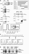
Localization of the homo-oligomerization domain with VP35 mutants. (A) Coimmunoprecipitation of VP35Flag and VP35 mutants. Flag-tagged VP35 was cotranslated with VP35 mutants with the TNT T7 Quick Coupled Reticulocyte Lysate System and metabolically labeled with [35S]methionine. In vitro translation was followed by coimmunoprecipitation with an anti-Flag or an anti-VP35 antibody. Immunocomplexes were separated by SDS-PAGE and visualized by Bio-Imager analysis. The positions of Flag-tagged VP35 and VP35 mutants are indicated. As a result of internal translation initiation in addition to the full-length product, smaller proteins were synthesized from the templates (*). A schematic representation of the MARV VP35 deletion mutants used for coimmunoprecipitation analyses with VP35Flag is shown at the upper right. The subscript numbers refer to the amino acids of VP35 in the respective mutants. A plus sign indicates an interaction with VP35Flag; a minus sign indicates no interaction with VP35Flag. (B) In silico analysis of the amino acid sequence of VP35 with the COILS 2.2 program. The graph shows a high probability of a coiled-coil structure between amino acids 70 and 120. (Insert) VP35Flag and the VP35 mutant lacking the potential coiled-coil domain (VP35Δ71-119) were cotranslated and analyzed for interaction by coimmunoprecipitation with a Flag-specific and an anti-VP35 antibody. (C) Amino acid sequence of the presumed coiled-coil domain. Note the occurrence of hydrophobic amino acid residues at the first (a) and fourth (d) positions of the heptad repeat (bold and underlined). Results of an in silico analysis of the VP35 substitution mutants (leucine 90 and/or 104 changed to alanine) with the COILS 2.2 program is shown at the bottom. (D) Flag-tagged VP35 substitution mutants were cotranslated with untagged VP35 substitution mutants and analyzed for interaction by coimmunoprecipitation as described above. The positions of the proteins are shown. As a result of internal translation initiation in addition to the full-length products, smaller proteins were synthesized from the templates (*).
FIG. 3.
FIG. 3.
The predicted coiled-coil domain of VP35 is sufficient to mediate homo-oligomerization. The putative homo-oligomerization domain spanning amino acids 70 to 120 was attached to a Flag-tagged (VP35-repFlag) and an untagged (VP35-rep) monomeric reporter protein. Leucine residues 90 and 104 within the homo-oligomerization domain of the generated constructs were replaced with alanine (mut-rep and mut-repFlag). The untagged monomeric reporter protein (rep) and the fusion proteins generated (VP35-rep, mut-rep) were coexpressed with their Flag-tagged versions, respectively. Complex formation was analyzed by coimmunoprecipitation with an anti-Flag monoclonal antibody and anti-VP30 rabbit immune serum. The proteins were separated by SDS-PAGE and visualized by Bio-Imager analysis. The positions of the proteins are indicated.
FIG. 4.
FIG. 4.
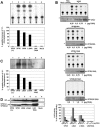
Influence of homo-oligomerization of VP35 on MARV transcription and replication. (A) Transcription analysis. Huh-T7 cells (5 × 10⁵) were transfected with plasmids encoding the MARV nucleocapsid proteins NP, L, and VP35 and VP35 substitution mutants. Additionally, a plasmid encoding the MARV-specific artificial minigenome 3 M-5 M and a plasmid expressing the T7 RNA polymerase were transfected. At 2 days posttransfection, cells were lysed and CAT activity was determined with [14C]chloramphenicol and acetyl coenzyme A as a substrate. The acetylated chloramphenicol was separated via thin-layer chromatography. Since chloramphenicol has two acetylation sites and the respective products have different migration velocities in thin-layer chromatography, two products can be distinguished beside the slow-migrating nonacetylated chloramphenicol when CAT activity is present. Quantification of CAT activity was done by the TINA 2.0 software (Raytest). (B) Titration of VP35 and VP35 substitution mutants. Huh-T7 cells were transfected as described above. The transfected amounts of VP35 or VP35 substitution mutants are indicated. Quantification of CAT activity was done by the TINA 2.0 software (Raytest). As the primary antibody for Western blotting, guinea pig anti-VP35 serum (dilution, 1:5,000) was used, and as the secondary antibody, a peroxidase-coupled anti-guinea pig antibody (DAKO; dilution, 1:25,000) was used. *, unspecific cellular proteins. Quantitative presentation of the titration of VP35, VP35L90A, and VP35L104A. (C) Replication analysis. Huh-T7 cells were transfected as described above. At 2 days posttransfection, cell lysates were treated with micrococcal nuclease. The isolated RNA was used to perform Northern blot analysis with digoxigenin-labeled riboprobes. Quantification of CAT activity was done by the TINA 2.0 software (Raytest). (D) Western blot assay. NP was detected by a mouse monoclonal anti-NP antibody (dilution, 1:1,000) and a peroxidase-coupled anti-mouse antibody (DAKO; dilution, 1:40,000). The expression levels of the respective mutants were checked by Western blot analysis. As the primary antibody, a guinea pig anti-VP35 serum (dilution, 1:5,000) was used, and as secondary antibody, a peroxidase-coupled anti-guinea pig antibody (DAKO; dilution, 1:25,000) was used.
FIG. 5.
FIG. 5.
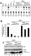
Impact of mixed oligomers derived from VP35 and VP35 truncation mutants on MARV transcription. (A) The minigenome system was set up in 5 × 10⁵ Huh-T7 cells as described in the legend to Fig. 4A. VP35 was replaced either with VP351-122 or VP351-122mut or with a mixture of VP35 (500 ng) and either VP351-122 or VP351-122mut in increasing amounts. A CAT assay was performed, and the radioactive signals were detected with a Bio-Imaging Analyzer. (B) Quantification of CAT activity was done by the TINA 2.0 software (Raytest). (C) Western blot assay. The expression levels of NP, VP35, and the respective mutants were checked by Western blot analysis. The antibodies used for detection of NP, VP35, and VP35 mutants are described in the legend to Fig. 4C.
FIG. 6.
FIG. 6.
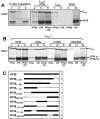
Influence of homo-oligomerization of VP35 on interaction with L. (A) LFlag and VP35 were coexpressed with the TNT T7 Quick Coupled Reticulocyte Lysate System and metabolically labeled with [35S]methionine. In vitro translation was followed by coimmunoprecipitation with an anti-Flag and/or an anti-VP35 antibody. Immunocomplexes were separated by SDS-PAGE and visualized by Bio-Imager analysis. To exclude antibody cross-reactivities, LFlag and VP35 were singly expressed in vitro and precipitated with the indicated antibodies. The positions of the proteins are indicated. (*) In addition to the full-length products, smaller proteins are synthesized from the templates as a result of internal translation initiation. (B) The untagged VP35 substitution mutants (leucine 90 and/or 104 changed to alanine) were cotranslated with LFlag and analyzed for interaction by coimmunoprecipitation as described above. The positions of the proteins are shown. (*) In addition to the full-length product, smaller proteins are synthesized from the templates as a result of internal translation initiation. (C) Schematic representation of the MARV VP35 deletion mutants used for coimmunoprecipitation analyses with LFlag. The subscript numbers refer to the amino acids of VP35 in the respective mutants. A plus sign indicates an interaction with LFlag; a minus sign indicates no interaction with LFlag.
FIG. 7.
FIG. 7.
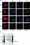
Influence of the homo-oligomerization of VP35 on colocalization of VP35 and NP-induced inclusions. (Top) VP35Flag or the Flag-tagged VP35 substitution mutants were either singly expressed or coexpressed together with NP. Immunofluorescence analysis was performed with a rabbit anti-Flag antibody (Sigma; dilution, 1:200) and a mouse monoclonal anti-NP antibody (dilution, 1:25). As secondary antibodies, a goat anti-rabbit immunoglobulin G antibody conjugated with rhodamine and a goat anti-mouse antibody conjugated with fluorescein isothiocyanate (Dianova, dilution, 1:200) were used. VP35Flag was expressed singly (A1) or coexpressed with NP (A2 to A4). (B to D) Single expression of the VP35 substitution mutant L90AFlag, L104AFlag, or L90/104AFlag (B1 to D1) versus coexpression together with NP (B2 to B4, C2 to C4, D2 to D4). (Bottom) Huh-T7 cells were transfected with plasmids encoding NP (100 ng) and VP35Flag or VP35 substitution mutant L90/104AFlag (500 ng) with Lipofectamine Plus (Invitrogen) according to the manufacturer's protocol. To exclude antibody cross-reactivities, NP was singly expressed and precipitated with the indicated antibodies. At 12 h posttransfection, cells were labeled with [35S]Promix and the proteins were immunoprecipitated with an anti-Flag antibody or an anti-NC serum, which is directed against the nucleocapsid proteins NP, VP35, and VP30. Immunocomplexes were separated by SDS-PAGE and visualized by Bio-Imager analysis. *, unspecific cellular proteins.

Similar articles

Cited by

References

    1. Asenjo, A., and N. Villanueva. 2000. Regulated but not constitutive human respiratory syncytial virus (HRSV) P protein phosphorylation is essential for oligomerization. FEBS Lett. 467:279-284. - PubMed
    1. Becker, S., and E. Mühlberger. 1999. Co- and posttranslational modifications and functions of Marburg virus proteins. Curr. Top. Microbiol. Immunol. 235:23-34. - PubMed
    1. Becker, S., C. Rinne, U. Hofsäss, H.-D. Klenk, and E. Mühlberger. 1998. Interactions of Marburg virus nucleocapsid proteins. Virology 249:406-417. - PubMed
    1. Centers for Disease Control and Prevention. 2005. Outbreak of Marburg virus hemorrhagic fever—Angola, October 1, 2004-March 29, 2005. Morb. Mortal. Wkly. Rep. 54:308-309. - PubMed
    1. Choudhary, S. K., A. G. Malur, Y. Huo, B. P. De, and A. K. Banerjee. 2002. Characterization of the oligomerization domain of the phosphoprotein of human parainfluenza virus type 3. Virology 302:373-382. - PubMed

Publication types

Substances

LinkOut - more resources