Lamellar organization of pigments in chlorosomes, the light harvesting complexes of green photosynthetic bacteria
- PMID: 15298919
- PMCID: PMC1304455
- DOI: 10.1529/biophysj.104.040956
Lamellar organization of pigments in chlorosomes, the light harvesting complexes of green photosynthetic bacteria
Abstract
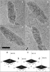
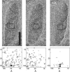
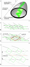
Chlorosomes of green photosynthetic bacteria constitute the most efficient light harvesting complexes found in nature. In addition, the chlorosome is the only known photosynthetic system where the majority of pigments (BChl) is not organized in pigment-protein complexes but instead is assembled into aggregates. Because of the unusual organization, the chlorosome structure has not been resolved and only models, in which BChl pigments were organized into large rods, were proposed on the basis of freeze-fracture electron microscopy and spectroscopic constraints. We have obtained the first high-resolution images of chlorosomes from the green sulfur bacterium Chlorobium tepidum by cryoelectron microscopy. Cryoelectron microscopy images revealed dense striations approximately 20 A apart. X-ray scattering from chlorosomes exhibited a feature with the same approximately 20 A spacing. No evidence for the rod models was obtained. The observed spacing and tilt-series cryoelectron microscopy projections are compatible with a lamellar model, in which BChl molecules aggregate into semicrystalline lateral arrays. The diffraction data further indicate that arrays are built from BChl dimers. The arrays form undulating lamellae, which, in turn, are held together by interdigitated esterifying alcohol tails, carotenoids, and lipids. The lamellar model is consistent with earlier spectroscopic data and provides insight into chlorosome self-assembly.
Figures


Similar articles
-
Internal structure of chlorosomes from brown-colored chlorobium species and the role of carotenoids in their assembly.Biophys J. 2006 Aug 15;91(4):1433-40. doi: 10.1529/biophysj.106.084228. Epub 2006 May 26. Biophys J. 2006. PMID: 16731553 Free PMC article.
-
X-ray scattering and electron cryomicroscopy study on the effect of carotenoid biosynthesis to the structure of Chlorobium tepidum chlorosomes.Biophys J. 2007 Jul 15;93(2):620-8. doi: 10.1529/biophysj.106.101444. Epub 2007 Apr 27. Biophys J. 2007. PMID: 17468163 Free PMC article.
-
Transmission electron microscopic study on supramolecular nanostructures of bacteriochlorophyll self-aggregates in chlorosomes of green photosynthetic bacteria.J Biosci Bioeng. 2006 Aug;102(2):118-23. doi: 10.1263/jbb.102.118. J Biosci Bioeng. 2006. PMID: 17027873
-
A model of the protein-pigment baseplate complex in chlorosomes of photosynthetic green bacteria.Photosynth Res. 2010 Jun;104(2-3):233-43. doi: 10.1007/s11120-009-9519-y. Epub 2010 Jan 14. Photosynth Res. 2010. PMID: 20077007 Review.
-
Chlorosome antenna complexes from green photosynthetic bacteria.Photosynth Res. 2013 Oct;116(2-3):315-31. doi: 10.1007/s11120-013-9869-3. Epub 2013 Jun 13. Photosynth Res. 2013. PMID: 23761131 Review.
Cited by
-
Anoxygenic photosynthesis with emphasis on green sulfur bacteria and a perspective for hydrogen sulfide detoxification of anoxic environments.Front Microbiol. 2024 Jul 11;15:1417714. doi: 10.3389/fmicb.2024.1417714. eCollection 2024. Front Microbiol. 2024. PMID: 39056005 Free PMC article. Review.
-
What We Are Learning from the Diverse Structures of the Homodimeric Type I Reaction Center-Photosystems of Anoxygenic Phototropic Bacteria.Biomolecules. 2024 Mar 6;14(3):311. doi: 10.3390/biom14030311. Biomolecules. 2024. PMID: 38540731 Free PMC article. Review.
-
Manifestation of Hydrogen Bonding and Exciton Delocalization on the Absorption and Two-Dimensional Electronic Spectra of Chlorosomes.J Phys Chem B. 2023 Feb 9;127(5):1097-1109. doi: 10.1021/acs.jpcb.2c07143. Epub 2023 Jan 25. J Phys Chem B. 2023. PMID: 36696537 Free PMC article.
-
Photosynthetic Light-Harvesting (Antenna) Complexes-Structures and Functions.Molecules. 2021 Jun 3;26(11):3378. doi: 10.3390/molecules26113378. Molecules. 2021. PMID: 34204994 Free PMC article. Review.
-
Superradiance of bacteriochlorophyll c aggregates in chlorosomes of green photosynthetic bacteria.Sci Rep. 2021 Apr 16;11(1):8354. doi: 10.1038/s41598-021-87664-3. Sci Rep. 2021. PMID: 33863954 Free PMC article.
References
-
- Balaban, T. S., A. R. Holzwarth, K. Schaffner, G. J. Boender, and H. J. de Groot. 1995. CP-MAS 13C-NMR dipolar correlation spectroscopy of 13C-enriched chlorosomes and isolated bacteriochlorophyll c aggregates of Chlorobium tepidum: The self organization of pigments is the main structural feature of chlorosomes. Biochemistry. 34:15259–15266. - PubMed
-
- Blankenship, R. E., J. M. Olson, and M. Miller. 1995. Antenna complexes from green photosynthetic bacteria. In Anoxygenic Photosynthetic Bacteria. R. E. Blankenship, M. T. Madigan, and C. E. Bauer, editors. Kluwer Academic Publisher, Dordrecht, The Netherlands. 399–435.
-
- Borrego, C. M., P. G. Gerola, M. Miller, and R. P. Cox. 1999. Light intensity effects on pigment composition and organisation in the green sulfur bacterium Chlorobium tepidum. Photosynth. Res. 59:159–166.
-
- Brune, D. C., G. H. King, and R. E. Blankenship. 1988. Interactions between bacteriochlorophyll c molecules in oligomers and in chlorosomes of green photosynthetic bacteria. In Photosynthetic Light-Harvesting Systems. H. Scheer,and S. Schneider, editors. Walter de Gruyter, Berlin. 141–151.
-
- Chiefari, J., K. Griebenow, F. Fages, N. Griebenow, T. S. Balaban, A. R. Holzwarth, and K. Schaffner. 1995. Models for the pigment organization in the chlorosomes of photosynthetic bacteria: Diastereoselective control of in vivo bacteriochlorophyll cs aggregation. J. Phys. Chem. 99:1357–1365.
Publication types
MeSH terms
Substances
LinkOut - more resources
Full Text Sources

