. 2024 Nov 19;40(1):100.
doi: 10.1007/s10565-024-09938-6.

The pivotal role of ZNF384: driving the malignant behavior of serous ovarian cancer cells via the LIN28B/UBD axis

Affiliations

The pivotal role of ZNF384: driving the malignant behavior of serous ovarian cancer cells via the LIN28B/UBD axis

Ye Yang et al. Cell Biol Toxicol. .

Abstract

Zinc finger protein 384 (ZNF384) is a highly conserved transcribed gene associated with the development of multiple tumors, however, its role and mechanism in serous ovarian cancer (SOC) are unknown. We first confirmed that ZNF384 was abnormally highly expressed in SOC tissues by bioinformatics analysis and immunohistochemistry. We further used lentivirus packaging and transfection techniques to construct ZNF384 overexpression or knockdown cell lines, and through a series of cell function experiments, gradually verified that ZNF384 promoted a series of malignant behaviors of SOC cell proliferation, migration, and invasion. By establishing a xenotransplantation model in nude mice, it was confirmed that ZNF384 promoted the progress of SOC in vivo. Mechanistically, Overexpression of ZNF384 enhanced the transcriptional activity of Lin-28 homolog B (LIN28B), which promoted the malignant behavior of SOC cells. In addition, LIN28B could regulate the expression of the downstream factor ubiquitin D (UBD) in SOC cells, further promoting the development of SOC. This study shows that ZNF384 aggravates the malignant behavior of SOC cells through the LIN28B/UBD axis, which may be used as a diagnostic biomarker for patients with SOC.

Keywords: LIN28B; Serous ovarian cancer; UBD; ZNF384.


Conflict of interest statement

Declarations Ethics approval All procedures involving human subjects were approved by Shengjing Hospital of China Medical University (Approval No. 2023PS426K), and animal experiments were performed with consent from the ethics committee of China Medical University (Approval No. CMU2023049). Consent for publication All authors have read the paper and agree that it can be published. Competing interests The authors declare no competing interests.

Figures

Fig. 1
Fig. 1
Identification of DEGs and their functional annotation in serous ovarian cancer. a GO and KEGG enrichment analysis of up-regulated DEGs in GSE14407 serous ovarian cancer database. Raw data between serous ovarian cancer and normal ovarian tissue were obtained from the Gene Expression Omnibus (GEO; http://www.ncbi.nlm.nih.gov/gds/) dataset and the gene expression profiling (GSE14407) was selected. Differentially expressed genes (DEGs) were selected by |log2FC|> 1 and adj. P < 0.05 in the GSE14407 dataset. Gene Ontology (GO) enrichment analysis and Kyoto Encyclopedia of Genes and Genomes (KEGG) enrichment analysis software were R packages ggplot2. b Enrichment fraction diagram of GO-GSEA in the GSE14407 dataset. c Enrichment fraction diagram of KEGG-GSEA in the GSE14407 dataset. Gene Set Enrichment Analysis (GSEA) analysis software was R package clusterProfiler + enrichplot. GO, Gene Ontology; KEGG, Kyoto Encyclopedia of Gene and Genome; DEG, differentially expressed genes; GSEA, Gene Set Enrichment Analysis
Fig. 2
Fig. 2
ZNF384 is highly expressed in serous ovarian cancer. a Circos plot showed the location of up-regulated genes in chromosomes of the GSE14407 dataset. The outermost track showed the chromosome information of these genes. The box plot in the middle of the Circos plot showed the expression of ZNF384 in normal and cancer tissue in the GSE14407 database. b Relative expression of ZNF384 in 30 serous ovarian cancer tissues and 30 normal ovarian tissues by qPCR. c The genomic profile of ZNF384 was obtained from the serous ovarian cancer data set (TCGA) using cBioportal database (https://www.cbioportal.org/). d The association between ZNF384 expression and OS, PFS, DFI, and DSS in serous ovarian cancer from the GSCA database (https://guolab.wchscu.cn/GSCA/). e Immunohistochemistry staining images of high and low expression of ZNF384. The scale bar indicates 50 µm. TCGA, The Cancer Genome Atlas; GSCA, Gene Set Cancer Analysis; OS, overall survival; PFS, progression-free survival; DFI, disease-free interval; DSS, disease-specific survival. ****p < 0.0001 as compared to the Normal group. Data are presented as mean ± SD. qPCR, quantitative real-time PCR
Fig. 3
Fig. 3
ZNF384 promotes the growth of serous ovarian cancer cells in vitro. a Western blot analysis of ZNF384 expression in normal ovarian epithelial cells (IOSE-80) and 5 lines of serous ovarian cancer cells (OVCAR-8, HEY A8, OVCAR-3, HEY and SKOV3). b Western blot and qPCR analysis of ZNF384 expression in OVCAR-8 and OVCAR-3 cells with ZNF384 knockdown. c Analysis of ZNF384 expression in SKOV3 cells with ZNF384 overexpression. ****p < 0.0001 as compared to the shCon or Vector group. d Cell proliferation of OVCAR-8, OVCAR-3, and SKOV3 cells after lentivirus infection was detected by CCK8 assay. *p < 0.05, **p < 0.01 as compared to the shCon or Vector group. e Colony formation assay of OVCAR-8, OVCAR-3, and SKOV3 cells was performed. The experiments were repeated three times. **p < 0.01, ***p < 0.001, ****p < 0.0001 as compared to the shCon or Vector group. Data are presented as mean ± SD. qPCR, quantitative real-time PCR
Fig. 4
Fig. 4
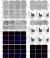
ZNF384 promotes the invasion and migration of serous ovarian cancer cells. a The effects of ZNF384 on the migration of serous ovarian cancer cells by wound healing assay. Photographs showed cell migration before and after injury under the microscope at 100 × magnification field. Quantification of cell migration by measuring wound closure areas before and after injury. b Effect of ZNF384 on the invasion of serous ovarian cancer cells by transwell assay (crystal violet staining × 200). c MMP-2 levels in the supernatants of OVCAR-8, OVCAR-3, and SKOV3 cells were detected by ELISA assay. *p < 0.05, **p < 0.01, ****p < 0.0001 as compared to the shCon or Vector group. d Representative immunofluorescence images of vimentin expression (red) in OVCAR-8, OVCAR-3, and SHOV3 cells. Cell nuclei were stained by DAPI (blue). The scale bar indicates 50 µm. Data are presented as mean ± SD. DAPI, 4′6’-diamidino-2-phenylindole dihydrochloride
Fig. 5
Fig. 5
ZNF384 promotes tumor growth and metastasis in vivo. a-c Xenograft tumors were formed by subcutaneous injection of OVCAR-8 or SKOV3 cells into BALB/c nude mice. The tumors were stripped after 25 days, photographed, measured, and weighed. *p < 0.05, **p < 0.01, ****p < 0.0001 as compared to the shCon or Vector group. d Immunohistochemical staining images of ZNF384, Ki67 expression in tumor tissues. The scale bar indicates 50 µm. e Bioluminescence images of BALB/c nude mice after intraperitoneal injection of OVCAR-8 or SKOV3 cells. f Representative images of abdominal cavity metastasis of tumors in different groups of nude mice (left panel) and quantification of the number of tumor metastatic nodules (right panel). ***p < 0.001, ****p < 0.0001 as compared to the shCon or Vector group. Data are presented as mean ± SD
Fig. 6
Fig. 6
ZNF384 promotes transcriptional activity of LIN28B in serous ovarian cancer. a-b Western blot and qPCR analysis of LIN28B expression in OVCAR-8, OVCAR-3, and SKOV3 cells with ZNF384 knockdown or overexpression. ***p < 0.001, ****p < 0.0001 as compared to the shCon or Vector group. c Predicted binding site and sequence names of LIN28B promoter (left panel). Dual-luciferase vectors containing LIN28B promoter sequences with different binding sites were co-transfected with ZNF384 overexpression plasmid into OVCAR-8 cells, and luciferase reporter gene assay was performed after 48 h (right panel). d The binding of ZNF384 to the LIN28B promoter was verified by ChIP experiments. e OVCAR-8 cells were transfected with LIN28B overexpression plasmid, and the transfection efficiency was verified by western blot and qPCR. f Cell proliferation of OVCAR-8 cells with LIN28B overexpression was detected by CCK8 assay. g-h The migration (g) and invasion (h) capacity of the OVCAR-8 cells transfected with LIN28B by wound healing and transwell assays. *p < 0.05, **p < 0.01. Data are presented as mean ± SD. qPCR, quantitative real-time PCR; ChIP, chromatin immunoprecipitation
Fig. 7
Fig. 7
LIN28B promotes the malignant behavior of serous ovarian cancer cells by regulating UBD. a Downstream factors that LIN28B may regulate were verified by RIP assay. SOX2 was used as a negative control. b Schematic diagram of predicted LIN28B motif and UBD binding sites. The binding of LIN28B and UBD was verified by RNA pull-down assay, and the expression of LIN28B in OVCAR-8 cells was detected by western blot. c Western blot and qPCR analysis of UBD expression in OVCAR-8 cells with LIN28B knockdown. d The half-life of UBD mRNA was detected in cells depicted in (c). e OVCAR-8 cells were transfected with UBD overexpression plasmid, and the transfection efficiency was verified by western blot and qPCR. Cell proliferation (f), migration (g), and invasion (h) ability of OVCAR-8 cells after transfection with UBD were detected by CCK-8, wound healing, and transwell assays, respectively. *p < 0.05, **p < 0.01, ***p < 0.001, ****p < 0.0001. Data are presented as mean ± SD. RIP, RNA-binding protein immunoprecipitation; qPCR, quantitative real-time PCR
Fig. 8
Fig. 8
Mechanism of ZNF384 promoting serous ovarian cancer progression. ZNF384 is involved in the malignant progression of serous ovarian cancer by promoting the proliferation, migration, and invasion of serous ovarian cancer cells. Its mechanism of action is related to the binding of the LIN28B promoter to promote its transcriptional activity, which further binds UBD mRNA, affects mRNA stability, and enhances its expression

Similar articles

References

    1. Boehm KM, Aherne EA, Ellenson L, Nikolovski I, Alghamdi M, Vázquez-García I, Zamarin D, Long Roche K, Liu Y, Patel D, et al. Multimodal data integration using machine learning improves risk stratification of high-grade serous ovarian cancer. Nat Cancer. 2022;3:723–33. 10.1038/s43018-022-00388-9. - PMC - PubMed
    1. Busch B, Bley N, Müller S, Glaß M, Misiak D, Lederer M, Vetter M, Strauß HG, Thomssen C, Hüttelmaier S. The oncogenic triangle of HMGA2, LIN28B and IGF2BP1 antagonizes tumor-suppressive actions of the let-7 family. Nucleic Acids Res. 2016;44:3845–64. 10.1093/nar/gkw099. - PMC - PubMed
    1. Chou CL, Chen TJ, Li WS, Lee SW, Yang CC, Tian YF, Lin CY, He HL, Wu HC, Shiue YL, et al. Upregulated ubiquitin D is a favorable prognostic indicator for rectal cancer patients undergoing preoperative concurrent chemoradiotherapy. Onco Targets Ther. 2022;15:1171–81. 10.2147/ott.S378666. - PMC - PubMed
    1. Cui P, Li H, Wang C, Liu Y, Zhang M, Yin Y, Sun Z, Wang Y, Chen X. UBE2T regulates epithelial-mesenchymal transition through the PI3K-AKT pathway and plays a carcinogenic role in ovarian cancer. J Ovarian Res. 2022;15:103. 10.1186/s13048-022-01034-9. - PMC - PubMed
    1. Dochez V, Caillon H, Vaucel E, Dimet J, Winer N, Ducarme G. Biomarkers and algorithms for diagnosis of ovarian cancer: CA125, HE4, RMI and ROMA, a review. J Ovarian Res. 2019;12:28. 10.1186/s13048-019-0503-7. - PMC - PubMed