

Basement Membranes in Development and Disease
- PMID: 29853176
- PMCID: PMC6701859
- DOI: 10.1016/bs.ctdb.2018.02.005
Basement Membranes in Development and Disease
Abstract
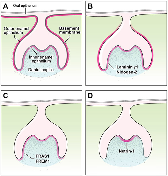
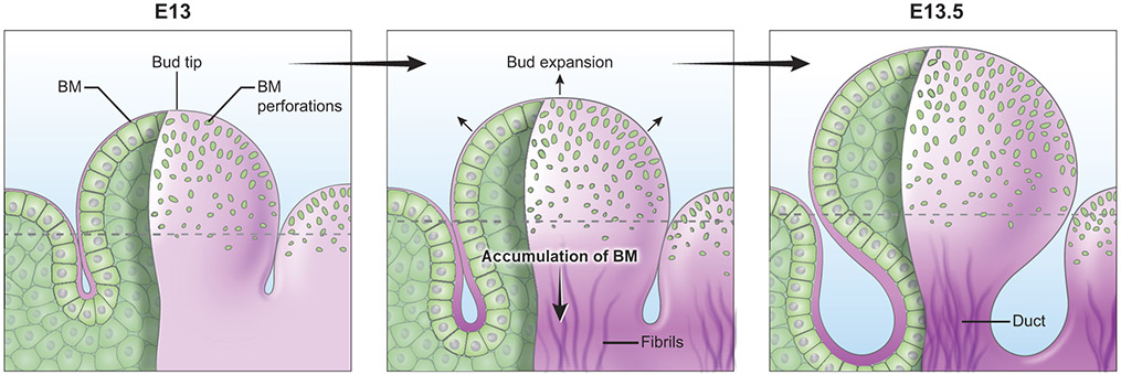
The basement membrane is a thin but dense, sheet-like specialized type of extracellular matrix that has remarkably diverse functions tailored to individual tissues and organs. Tightly controlled spatial and temporal changes in its composition and structure contribute to the diversity of basement membrane functions. These different basement membranes undergo dynamic transformations throughout animal life, most notably during development. Numerous developmental mechanisms are regulated or mediated by basement membranes, often by a combination of molecular and mechanical processes. A particularly important process involves cell transmigration through a basement membrane because of its link to cell invasion in disease. While developmental and disease processes share some similarities, what clearly distinguishes the two is dysregulation of cells and extracellular matrices in disease. With its relevance to many developmental and disease processes, the basement membrane is a vitally important area of research that may provide novel insights into biological mechanisms and development of innovative therapeutic approaches. Here we present a review of developmental and disease dynamics of basement membranes in Caenorhabditis elegans, Drosophila, and vertebrates.
Keywords: Basement membrane; Basement membrane pores; Cancer; Cell migration; Development; Extracellular matrix; Invasion; Morphogenesis; Proliferation.
2018 Published by Elsevier Inc.
Figures


Similar articles
-
Basement Membranes in the Worm: A Dynamic Scaffolding that Instructs Cellular Behaviors and Shapes Tissues.Curr Top Membr. 2015;76:337-71. doi: 10.1016/bs.ctm.2015.08.001. Epub 2015 Sep 12. Curr Top Membr. 2015. PMID: 26610919 Free PMC article. Review.
-
Basement membranes.Curr Biol. 2017 Mar 20;27(6):R207-R211. doi: 10.1016/j.cub.2017.02.006. Curr Biol. 2017. PMID: 28324731
-
A developmental biologist's "outside-the-cell" thinking.J Cell Biol. 2015 Aug 3;210(3):369-72. doi: 10.1083/jcb.201501083. J Cell Biol. 2015. PMID: 26240181 Free PMC article.
-
Heterotypic control of basement membrane dynamics during branching morphogenesis.Dev Biol. 2015 May 1;401(1):103-9. doi: 10.1016/j.ydbio.2014.12.011. Epub 2014 Dec 16. Dev Biol. 2015. PMID: 25527075 Free PMC article. Review.
-
Basement membranes.WormBook. 2005 Sep 1:1-15. doi: 10.1895/wormbook.1.16.1. WormBook. 2005. PMID: 18050423 Free PMC article. Review.
Cited by
-
CBD-1 organizes two independent complexes required for eggshell vitelline layer formation and egg activation in C. elegans.Dev Biol. 2018 Oct 15;442(2):288-300. doi: 10.1016/j.ydbio.2018.08.005. Epub 2018 Aug 16. Dev Biol. 2018. PMID: 30120927 Free PMC article.
-
Silencing TRIP13 inhibits cell growth and metastasis of hepatocellular carcinoma by activating of TGF-β1/smad3.Cancer Cell Int. 2018 Dec 17;18:208. doi: 10.1186/s12935-018-0704-y. eCollection 2018. Cancer Cell Int. 2018. PMID: 30564064 Free PMC article.
-
Ultrasonographic Assessment of the Cutaneous Changes Induced by Topical Use of Novel Peptides Comprising Laminin 5.Arch Plast Surg. 2022 May 27;49(3):304-309. doi: 10.1055/s-0042-1748642. eCollection 2022 May. Arch Plast Surg. 2022. PMID: 35832163 Free PMC article.
-
Cell-extracellular matrix dynamics.Phys Biol. 2022 Jan 12;19(2):10.1088/1478-3975/ac4390. doi: 10.1088/1478-3975/ac4390. Phys Biol. 2022. PMID: 34911051 Free PMC article. Review.
-
A chloride ring is an ancient evolutionary innovation mediating the assembly of the collagen IV scaffold of basement membranes.J Biol Chem. 2019 May 17;294(20):7968-7981. doi: 10.1074/jbc.RA119.007426. Epub 2019 Mar 28. J Biol Chem. 2019. PMID: 30923125 Free PMC article.
References
-
- Aumailley M, Bruckner-Tuderman L, Carter WG, Deutzmann R, Edgar D, Ekblom P, Engel J, Engvall E, Hohenester E, Jones JC, Kleinman HK, Marinkovich MP, Martin GR, Mayer U, Meneguzzi G, Miner JH, Miyazaki K, Patarroyo M, Paulsson M, Quaranta V, Sanes JR, Sasaki T, Sekiguchi K, Sorokin LM, Talts JF, Tryggvason K, Uitto J, Virtanen I, von der Mark K, Wewer UM, Yamada Y, Yurchenco PD, 2005. A simplified laminin nomenclature. Matrix Biol 24, 326–332. - PubMed
Publication types
MeSH terms
Grants and funding
LinkOut - more resources
Full Text Sources
Other Literature Sources
Molecular Biology Databases
