Central tegmental tract: Difference between revisions
added ref |
→Structure: added to and ref |
||
| Line 14: | Line 14: | ||
==Structure== |
==Structure== |
||
The central tegmental tract contains descending and ascending fibers. It runs from the [[red nucleus]] to the inferior [[olivary nucleus]].<ref name="Standring2-2020">{{Cite book |last=Standring |first=Susan |url=https://www.worldcat.org/oclc/1201341621 |title=Gray's Anatomy: The Anatomical Basis of Clinical Practice |publisher=[[Elsevier]] |year=2020 |isbn=978-0-7020-7707-4 |edition=42nd |location=New York |pages=456–458 |oclc=1201341621}}</ref> |
The central tegmental tract contains descending and ascending fibers. It runs from the [[red nucleus]] to the inferior [[olivary nucleus]].<ref name="Standring2-2020">{{Cite book |last=Standring |first=Susan |url=https://www.worldcat.org/oclc/1201341621 |title=Gray's Anatomy: The Anatomical Basis of Clinical Practice |publisher=[[Elsevier]] |year=2020 |isbn=978-0-7020-7707-4 |edition=42nd |location=New York |pages=456–458 |oclc=1201341621}}</ref> |
||
===Descending fibers=== |
===Descending fibers=== |
||
Latest revision as of 07:53, 31 October 2024
| Central tegmental tract | |
|---|---|
 Diagram of the midbrain, sectioned at the level of the superior colliculus (Central tegmental tract not labeled, but region is visible.) | |
 Axial section of the Brainstem (Pons) at the level of the Facial Colliculus (Central tegmental tract not labeled, but region is visible.) | |
| Details | |
| Identifiers | |
| Latin | Tractus tegmentalis centralis |
| NeuroNames | 2204 |
| TA98 | A14.1.05.325 |
| TA2 | 5869 |
| FMA | 83850 |
| Anatomical terms of neuroanatomy | |
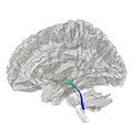
The central tegmental tract is a tract that carries ascending and descending fibers, situated in the midbrain tegmentum, and the pontine tegmentum. The tract is situated in the central portion of the reticular formation.[1]
Structure
[edit]The central tegmental tract contains descending and ascending fibers. It runs from the red nucleus to the inferior olivary nucleus.[2] In humans it is very small and runs ventral to the lateral corticospinal tract.[2]
Descending fibers
[edit]Descending fibers of the rubro-olivary tract project from the parvocellular red nucleus to the ipsilateral inferior olivary nucleus.[1][3] The inferior olivary nucleus projects to the contralateral cerebellum via olivocerebellar fibers.[3] The rubro-olivary fibres descend through the superior cerebellar peduncle.[1]
Ascending fibers
[edit]Ascending fibers are second-order axons projecting from the gustatory nucleus (the rostral part of the solitary nucleus] to the ventral posteromedial nucleus of thalamus[1][3] (third-order neurons in turn project to the gustatory cortex).
Ascending reticulothalamic fibres project from the medial zone nuclei of the reticular formation to the hypothalamus (to mediate autonomic nervous system response), and the intralaminar thalamic nuclei (to mediate a startle response to pain).[3]
Clinical significance
[edit]Lesion of the tract can cause palatal myoclonus, e.g. in myoclonic syndrome, in strokes of the posterior inferior cerebellar artery.[citation needed]
Additional Images
[edit]-
Horizontal section through the lower part of the pons. The central tegmental tract is labeled #16.
-
Tractography showing central tegmental tract
References
[edit]- ^ a b c d Standring, Susan (2020). Gray's Anatomy: The Anatomical Basis of Clinical Practice (42th ed.). New York: Elsevier. p. 451. ISBN 978-0-7020-7707-4. OCLC 1201341621.
- ^ a b Standring, Susan (2020). Gray's Anatomy: The Anatomical Basis of Clinical Practice (42nd ed.). New York: Elsevier. pp. 456–458. ISBN 978-0-7020-7707-4. OCLC 1201341621.
- ^ a b c d Patestas, Maria A.; Gartner, Leslie P. (2016). A Textbook of Neuroanatomy (2nd ed.). Hoboken, New Jersey: Wiley-Blackwell. pp. 112, 292, 298, 306, 473. ISBN 978-1-118-67746-9.
External links
[edit]- Midbrain at Inferior Colliculus - IV Nucleus, Sectional Atlas
- Mid Pons at the Trigeminal Motor Nucleus, Sectional Atlas
- Neuroanatomy / plate12, Frank Willard


