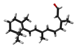
Retinal

| |
Except where otherwise noted, data are given for materials in their standard state (at 25 °C [77 °F], 100 kPa).
|
Retinal, technically called retinene1 or retinaldehyde, is a light-sensitive retinene molecule found in the photoreceptor cells of the retina. Retinal is the fundamental chromophore involved in the transduction of light into visual signals, i.e. nerve impulses, in the visual system of the central nervous system.
Overview
The molecule that takes part in the initial step in the vision process, rhodopsin, has two components called 11-cis retinal and opsin. Retinal is a light-sensitive derivative of vitamin A, and opsin is a protein molecule. Rhodopsin is found in the rod cells of the eye. 11-cis retinal is a powerful absorber of light because it is a polyene; its 6 alternating single and double bonds make up a long conjugated electron network. When no light is present, the 11-cis retinal molecule is found in a "bent (cis) configuration" (fig A), and as such it is attached to the opsin molecule in a stable arrangement:

When light strikes the retina, a retinal molecule may absorb a photon, promoting it into an excited electronic state. The nature of the excited state is not well understood, but it is known that within 200 femtoseconds it returns to the ground electronic state.[1] One third of these events cause no net change, while the remaining two thirds induce a rotation in the pi bond found between the eleventh and twelfth carbon atoms. In other words, the 11-cis retinal is transformed into the all-trans retinal (fig B) in a straightened configuration.[2]
It takes only one rhodopsin isomerization to reliably generate a rod electrical signal.[3][4] In mammals, a single rod photon absorption will lead ganglion cells at the output layer of the retina to fire 2-3 additional action potentials,[5] although it may require additional photon absorptions before this signal is detectable above the ganglion cell baseline noise.
The all-trans retinal configuration, subsequently, does not fit into the binding site of the opsin molecule in the same manner as the 11-cis configuration; as a result, upon isomerization, the trans isomer causes a conformational change in the protein, which triggers a G protein signaling pathway' including transducin, that results in the generation of an electrical impulse, which is transmitted through the optic nerve to the brain for processing. Trans retinal is eventually removed from opsin, and then recycled back to the 11-cis isomer for subsequent use. Enzymes mediate the isomerization of all-trans back to the 11-cis configuration, and rhodopsin is regenerated by a new formation of a Schiff base linkage, which actuates the binding of the cis isomer to opsin. This is the basic mechanism of the vision cycle.
All-trans-retinal is also an essential component of type I, or microbial, opsins such as bacteriorhodopsin, channelrhodopsin, and halorhodopsin. In these molecules, light causes the all-trans-retinal to become 13-cis retinal,[6] which then cycles back to all-trans-retinal in the dark state.
 |
 |
History
This photon induced retinal-bending mechanism was discovered in 1958 by the American biochemist George Wald and his co-workers. For his work, Wald won a share of the 1967 Nobel Prize in Physiology or Medicine with Haldan Keffer Hartline and Ragnar Granit.[7]
See also
References
- ^ van Dorst, W.C.A. (28 Januari 1987). "Quantumchemische berekeningen aan retinal-modelstoffen" (in Dutch). Eindhoven University of Technology, Faculty of Chemical Technology, Organic Chemistry dept: 35 pages.
{{cite journal}}: Check date values in:|date=(help); Cite journal requires|journal=(help); Unknown parameter|coauthors=ignored (|author=suggested) (help) - ^ Chang, Raymond (1998). Chemistry, 6th Ed. New York: McGraw Hill. ISBN 0-07-115221-0. OCLC 60178489.
- ^ D. A. Baylor, T. D. Lamb, and K.-W. Yau. Responses of retinal rods to single photons. J Physiol, 288:613–634, March 1979.
- ^ S. Hecht, S. Shlaer, and M. H. Pirenne. Energy, quanta, and vision. The Journal of General Physiology, 25(6):819–840, 1942.
- ^ H. B. Barlow, W. R. Levick, and M. Yoon. Responses to single quanta of light in retinal ganglion cells of the cat. Vision Res, Suppl 3:87–101, 1971.
- ^ De-liang Chen, Guang-yu Wang, Bing Xu and Kun-sheng Hu. All-trans to 13-cis retinal isomerization in light-adapted bacteriorhodopsin at acidic pH. Journal of Photochemistry and Photobiology B:Biology. 2002 Apr; 66(3):188-94. doi:10.1016/S1011-1344(02)00245-2
- ^ 1967 Nobel Prize in Medicine
External links
- First Steps of Vision - National Health Museum
- Vision and Light-Induced Molecular Changes
- Retinal Anatomy and Visual Capacities
- Retinal
